Vadzo Imaging Positions Innova-1335CRA: AR1335 13MP Autofocus GigE Camera with Ultra Low Light Imaging and Digital PTZ for Pathology Devices
Built for digital pathology scanners and microscopy platforms that must resolve fine tissue detail under attenuated
Press Release Disclaimer: This is a press release distributed through the XPR Media network. It has not been independently verified by our newsroom.


Built for digital pathology scanners and microscopy platforms that must resolve fine tissue detail under attenuated illumination, this camera pairs the Onsemi AR1335 sensor with ultra-low-light sensitivity, digital PTZ, and autofocus over GigE Vision, giving pathology device manufacturers detailed 13MP imaging without a mechanical pan-tilt-zoom assembly or a proprietary interface to manage.
IRVINE, CA / ACCESS Newswire / August 24, 2026 / Vadzo Imaging, a world leader in embedded vision solutions, today announces the positioning of Innova-1335CRA, a 13MP Autofocus GigE Camera built around the Onsemi AR1335 sensor for digital pathology, whole slide imaging, and clinical microscopy applications that need ultra-low light sensitivity and standardized network connectivity.
Why Pathology Imaging Needs a 13MP Autofocus GigE Camera Built for Low Light Precision
Digital pathology imaging presents a lighting problem that most other camera applications never encounter in the same way. Many pathology and histology imaging workflows deliberately use attenuated illumination to avoid photobleaching a stained sample or to accommodate a thin tissue section that transmits only a fraction of incident light, which leaves a standard sensor working with far less usable signal than a typical machine vision or security application would ever provide. A camera without genuine ultra-low-light sensitivity forces a lab to choose between increasing illumination in ways that can degrade a sample and accepting noisy, unusable imagery.
An Ultra-Low-Light GigE Camera approaches that problem directly at the sensor level, extending usable sensitivity well past the point where a standard sensor would need brighter illumination to produce a clean image. This matters directly for stained tissue samples and transmitted light microscopy, where the sample being examined and the image quality needed to examine it are not competing priorities a lab should have to balance against each other.
A second, related challenge in pathology imaging is navigating a slide without introducing mechanical complexity or vibration into the optical path. A Digital PTZ Vision Camera solves that problem electronically, panning, tilting, and zooming within the sensor’s captured field through cropping and scaling rather than physically moving a lens assembly, which removes an entire category of mechanical wear and vibration risk from a slide scanner’s design.
These two challenges, limited light and navigation without mechanical complexity, tend to compound each other in a real pathology workflow. A scanner that solves ultra-low-light sensitivity but still relies on a mechanical PTZ mechanism introduces vibration risk during exactly the low-light, long-exposure captures where that vibration matters most. Addressing both from the same sensor platform gives a pathology device manufacturer one specification to evaluate rather than two separate engineering compromises pulling in different directions.
Engineering Explanation: Sensitivity, Navigation, and Focus Together
Onsemi designed the AR1335 as a 13-megapixel sensor capable of both full-resolution still capture and cropped video output, and Vadzo Imaging pairs that sensor with ultra-low-light optimization specifically for pathology and microscopy applications. As an AR1335 Autofocus GigE Camera, the sensor extends usable sensitivity into lighting conditions where a standard sensor would need substantially brighter illumination, which matters directly for stained tissue samples and transmitted light microscopy where excessive illumination can degrade the very sample being examined.
That sensitivity works alongside a second capability built specifically for slide navigation. As an AR1335 Low Light GigE Sensor, the Innova-1335CRA supports AR1335 PTZ Camera functionality entirely in the digital domain, cropping and scaling within the sensor’s captured field to pan, tilt, and zoom without a mechanical gimbal or lens assembly. A PTZ GigE Camera Module configuration like this one removes the vibration, wear, and calibration drift that a mechanical PTZ mechanism would introduce into a precision slide scanning workflow.
Autofocus adds a third dimension to this design that matters specifically for pathology samples. As an AR1335 Digital Zoom Camera, the Innova-1335CRA adjusts focus electronically as it navigates across a slide, which is necessary because tissue sections and mounting media rarely present a perfectly flat, uniform focal plane across an entire specimen. A Digital Zoom Ethernet Camera configuration keeps that focus accurate even as digital zoom changes the effective magnification of the region under examination.
Delivering all of this over GigE Vision is the final piece of the design. As a 13MP Gigabit Ethernet Camera, the Innova-1335CRA connects using a standard, GenICam-compliant protocol that most scientific imaging and pathology software platforms already support, and functioning as a 13MP Ultra Low Light Camera, it does so while running standard Ethernet cabling that most lab environments already have in place rather than requiring a specialized interface.
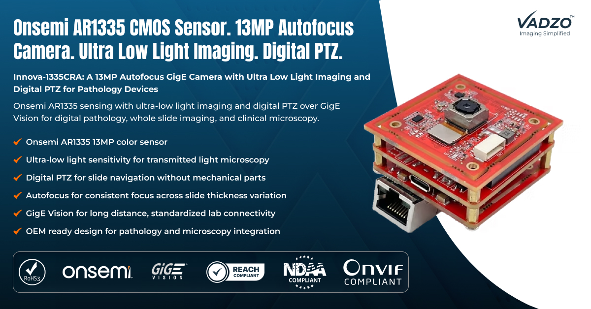
Product Overview
The Innova-1335CRA pairs the Onsemi AR1335 Pathology Camera sensor with ultra-low-light sensitivity, digital PTZ, and autofocus in a compact design suited to slide scanner, microscope, and pathology workstation integration. As a 13MP Autofocus GigE Camera, it outputs full 13-megapixel resolution with autofocus and ultra-low-light processing active by default, giving pathology device manufacturers a single camera platform that covers whole slide imaging, histology review, and general clinical microscopy from one module.
At the core of the module sits the AR1335 gigabit ethernet Camera sensor, chosen specifically for the combination of resolution, low-light sensitivity, and digital PTZ flexibility that pathology imaging requires. Because the camera pairs 4K Ultra Low Light Ethernet camera sensitivity with 13MP Digital Zoom Sensor navigation, the Innova-1335CRA reaches usable image quality across attenuated lighting conditions while giving lab technicians the ability to navigate a slide electronically rather than through mechanical stage or lens movement.
Product Specifications
Key specs: 13MP (4208 × 3120) Max Resolution | Onsemi AR1335, 1/3.2 inch Sensor Format | 1.1 µm Pixel | Color, Rolling Shutter Chroma and Shutter | GigE Vision (Gigabit Ethernet) | Autofocus Lens Assembly Optics
Key Capabilities
Onsemi AR1335 Sensor for Detailed 13MP Pathology Capture: The Innova-1335CRA is built around the Onsemi AR1335 sensor, a 13 megapixel device capable of resolving fine tissue and cellular detail across a full slide or a closely examined region. This gives the module genuine 13MP PTZ Camera Module performance suited to pathology applications that need both wide-field slide overview and detailed close examination from the same hardware. A pathologist or lab technician reviewing a slide typically needs to move fluidly between a wide overview and a closely examined region of interest, and a sensor that resolves both levels of detail from the same capture removes the need to switch between separate wide-field and close-up imaging hardware mid-review.
Ultra Low Light Imaging for Transmitted Light Microscopy: Stained tissue samples and thin sections examined under transmitted light often present far less usable signal than a typical machine vision application, and increasing illumination to compensate risks degrading the sample itself. As an Ultra Low Light Ethernet Camera, the Innova-1335CRA extends sensitivity well past the point where a standard sensor would require brighter illumination, preserving both sample integrity and image usability.
Digital PTZ for Slide Navigation Without Mechanical Parts: A mechanical pan-tilt-zoom assembly introduces vibration, wear, and calibration drift into a precision imaging workflow, none of which a digital pathology system can afford during a diagnostic review. As a Low Light Autofocus GigE Camera, the Innova-1335CRA navigates a slide entirely within the digital domain, cropping and scaling the sensor’s captured field to pan, tilt, and zoom without moving a single mechanical part. Because there is no motor, gimbal, or moving lens assembly involved in that navigation, there is also no mechanical component to wear out, drift out of calibration, or require periodic service over the equipment’s operating life, which matters directly for a diagnostic instrument expected to remain accurate across years of continuous laboratory use.
Autofocus for Consistent Focus Across Slide Thickness Variation: Tissue sections and mounting media rarely present a perfectly flat focal plane across an entire slide, which makes autofocus a genuine clinical requirement rather than a convenience feature for pathology imaging. The Innova-1335CRA’s autofocus lens assembly adjusts electronically as it navigates across a slide, keeping the region under examination sharp regardless of minor thickness variation across the specimen.
GigE Vision for Long Distance, Standardized Lab Connectivity: Most pathology and scientific imaging software already supports the GigE Vision and GenICam standards natively, which makes a GigE-connected camera a more direct fit for lab integration than a camera requiring a proprietary driver or capture card. As a GigE Low Light Camera Kit, the Innova-1335CRA connects using standard Ethernet cabling already common in lab environments, reaching workstations and network storage without a specialized interface. That standard connectivity also means a lab can position the camera at a meaningful distance from its workstation or network switch without the cable length restrictions a USB connection would introduce, which offers more flexibility in how a scanner or microscope station is physically laid out within a lab.
OEM Ready Design for Pathology and Microscopy Integration: Pathology scanner and microscopy equipment manufacturers producing units for clinical and research use need a camera platform that stays consistent across a long production run. Vadzo Imaging supports full customization on this platform as an OEM Pathology Camera Module, including connector changes, optics selection, and firmware adjustments for manufacturers headed into volume production. That level of customization extends to firmware behavior as well, since a manufacturer building a high-throughput screening platform may need different digital PTZ responsiveness than one building a detailed diagnostic review station, even though both use the same underlying sensor and connectivity.
“Pathology labs cannot afford a camera that forces a choice between illuminating a sample brightly enough to see clearly and preserving that same sample for further examination. The AR1335’s ultra-low light sensitivity combined with digital PTZ gives the Innova-1335CRA genuine diagnostic image quality without a mechanical pan-tilt-zoom assembly to maintain, delivered over standard GigE Vision networking that fits directly into existing lab infrastructure. We built this camera for workflows where the sample matters as much as the image.” – Alwin Vincent, Product Manager at Vadzo Imaging.
Application-Specific Sections
Digital Pathology and Whole Slide Imaging: Whole slide imaging systems need to resolve fine cellular detail across an entire specimen while navigating efficiently between regions of interest. As a Digital Pathology Camera, the Innova-1335CRA supports that complete workflow, and its digital PTZ handles navigation without mechanical movement. Functioning as a Pathology Slide Scanner Camera, it delivers the resolution and low light sensitivity a full slide scan actually requires, and as a Whole Slide Imaging Camera, it covers the complete specimen review process from initial scan to detailed regional examination. That end-to-end coverage from a single camera platform simplifies both the hardware a scanner manufacturer needs to qualify and the workflow a pathologist follows when moving between an overview scan and a detailed regional review of the same specimen.
Histology and Tissue Sample Imaging: Histology imaging depends on resolving fine tissue structure under staining and illumination conditions that vary meaningfully from one sample preparation to another. As a Histology Imaging Camera, the Innova-1335CRA’s ultra-low light sensitivity holds up across that variation, and a Pathology Imaging Camera Module configuration of the same module extends the same reliability to broader tissue sample review workflows.
Clinical Pathology Lab Integration: Clinical pathology labs running diagnostic workflows need imaging equipment that integrates directly with existing lab information systems and network infrastructure. As a Clinical Pathology Camera, the Innova-1335CRA’s GigE Vision connectivity fits that existing infrastructure, and functioning as a Lab Pathology Vision Camera, it supports both diagnostic imaging and general lab documentation tasks from the same hardware.
Research and Academic Microscopy: Research and academic microscopy applications often need to capture detailed imagery across a wide range of sample types and lighting conditions without the budget for dedicated hardware per application. A Microscopy gigabit Ethernet camera platform like this one gives research labs that flexibility, and as a GigE Vision Pathology Camera, it integrates directly with standard scientific imaging software already common in academic settings.
OEM Integration for Pathology Scanner Manufacturers: Pathology scanner and microscopy equipment manufacturers building their own diagnostic platforms need a camera vendor who understands the low light, navigation, and connectivity requirements those systems demand. The Innova-1335CRA ships from a PTZ Camera Manufacturer with those requirements built in, and Vadzo Imaging supports OEM customization for optics, connector configuration, and firmware for manufacturers standardizing on one camera across multiple pathology or microscopy product lines. As a Pathology Scanner Vision Module, it fits directly into a manufacturer’s existing platform architecture.
Frequently Asked Questions
Q: Why does a pathology imaging camera need ultra-low light sensitivity when lab lighting can simply be increased?
A: Many pathology imaging workflows deliberately use attenuated illumination rather than bright lighting, either because a stained sample can photobleach under intense light or because a thin tissue section transmits only a small fraction of incident light regardless of how bright the source is. Simply increasing illumination is not always an option without risking damage to the very sample being examined, which places the burden on the camera to extract a usable image from limited available light rather than on the lighting system to provide more of it. Vadzo Imaging builds ultra-low-light sensitivity into its pathology imaging camera products specifically because sample preservation and image quality cannot be treated as competing priorities in a diagnostic workflow.
Q: What is digital PTZ, and why would a slide scanner use it instead of a mechanical pan-tilt-zoom camera?
A: Digital PTZ achieves pan, tilt, and zoom electronically by cropping and scaling within a sensor’s already captured field of view, rather than physically moving a lens assembly or camera housing to change the framing. A mechanical pan-tilt-zoom mechanism introduces moving parts that can wear, drift out of calibration, and introduce vibration into a precision optical path, all of which matter more in a diagnostic slide scanning workflow than in a general security or surveillance application. Vadzo Imaging includes digital PTZ on its pathology imaging camera products so slide scanner manufacturers get flexible navigation without adding a mechanical subsystem that requires its own maintenance and calibration over the life of the equipment.
Q: Why would a pathology imaging system use a GigE Vision camera instead of a USB camera?
A: Most established pathology and scientific imaging software platforms already support the GigE Vision and GenICam standards natively, since these standards have been widely adopted across the broader machine vision and scientific imaging industry for years. A GigE Vision camera plugs directly into that existing software ecosystem without a proprietary driver, while also supporting longer cable runs over standard Ethernet infrastructure than a typical USB connection allows. Vadzo Imaging offers its pathology imaging camera products with GigE Vision connectivity specifically because it matches the standard most lab software and network infrastructure already expects, rather than requiring a specialized interface layer. That compatibility also simplifies long-term support, since a lab’s IT staff can rely on the same GigE Vision troubleshooting knowledge and network infrastructure across multiple pieces of imaging equipment from different vendors rather than learning a separate proprietary system for each device.
Q: Can Vadzo Imaging customize a pathology imaging camera module for a specific slide scanner or microscopy platform?
A: Yes. Vadzo Imaging supports full OEM customization across its pathology imaging camera portfolio, including optics selection suited to a specific magnification range or working distance, connector and cable configuration matched to a particular scanner or microscope design, and firmware adjustments tuned for a specific digital PTZ or low light sensitivity profile. Evaluation units ship with no minimum order requirement, and the same engineering team that built the standard product works directly with pathology and microscopy equipment manufacturers to adapt the module for a specific diagnostic or research platform headed into volume production. That level of customization extends to firmware behavior as well, since a manufacturer building a high-throughput screening platform may need different digital PTZ responsiveness than one building a detailed diagnostic review station, even though both use the same underlying sensor and connectivity.
Q: What should pathology and microscopy equipment manufacturers look for when choosing a camera supplier for a diagnostic imaging platform?
A: A diagnostic imaging platform depends on consistent image quality across every unit in a production run, since a scanner that performs inconsistently between units undermines confidence in the diagnostic results it produces. Manufacturers should look for genuine low-light sensitivity validated under realistic sample conditions rather than only bright bench test lighting, a connectivity standard already supported by established lab software, and a supplier able to maintain consistent hardware and firmware across a long product lifecycle. Vadzo Imaging builds its pathology imaging camera products around exactly these priorities, since a camera that performs well in a demo but varies unit to unit is not actually suitable for a diagnostic platform used in clinical or research settings. Requesting performance data across multiple evaluation units, rather than relying on a single sample unit’s specifications, is generally the more reliable way to confirm that consistency before committing to a volume order.
Availability
The Innova-1335CRA 13MP Autofocus GigE Camera is available now for evaluation and OEM integration. Evaluation kits include the camera module, a factory-calibrated autofocus lens, a GigE Vision cable, and integration documentation covering digital PTZ configuration and ultra-low-light tuning, with no minimum order requirement. Contact Vadzo Imaging at support@vadzoimaging.com to request an evaluation unit or discuss volume production and OEM customization. Evaluation feedback is also welcome directly through the same contact channel, since real laboratory deployment conditions often surface details a bench test alone would not reveal.
About Vadzo Imaging
Vadzo Imaging develops embedded and machine vision camera products for OEMs, system integrators, and pathology equipment manufacturers building production-ready vision systems across digital pathology, clinical diagnostics, industrial automation, and edge AI. The company’s portfolio spans MIPI CSI-2, USB, GigE, Wi-Fi, FPD-Link III, and SerDes interface camera products, supporting deployment architectures from compact onboard modules to distributed networked installations. Vadzo provides end-to-end imaging support, including sensor integration, ISP tuning, firmware development, and SDK frameworks to accelerate system deployment, and the company’s engineering team works directly with customers throughout evaluation, pilot deployment, and volume production rather than handing off support after the initial sale. That ongoing engineering relationship matters most for diagnostic and clinical imaging programs, where a camera platform may remain in service across a laboratory’s equipment fleet for many years after its initial installation. Visit Vadzo Imaging to explore the full embedded vision camera portfolio.
Media Contact
Alwin Vincent
Vadzo Imaging
Email: alwin@vadzoimaging.com
LinkedIn: Vadzo Imaging
YouTube: Vadzo Imaging
X: Vadzo Imaging
SOURCE: Vadzo Imaging
View the original press release on ACCESS Newswire
Media gallery